What Is It?
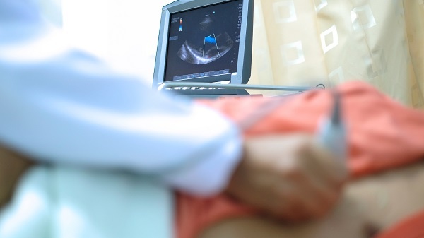
It is an ultrasound examination of the heart, showing the heart structure and function in real time.
How Is It Done?
A probe is placed on the chest in number of positions and ultrasound (sound wave) energy is used to produce pictures of the heart.
Why Is It Done?
Echocardiography shows whether the heart is structurally normal and functioning normally. It can show any heart chamber enlargement and abnormal heart function. The heart valves can be seen and assessed as to whether they are working properly. Any abnormal cardiac masses can be assessed as well as whether there is any fluid present in the sac that the heart sits in.
Echocardiograph gives accurate and immediate information on issues of heart function and structure. It is easily accessible and portable.